Transvaginal Ultrasound (TVS) in Malaysia: A Simple Guide for Women and Couples
A transvaginal ultrasound (TVS)—also called an endovaginal scan—is a safe and more reliable to examine the uterus, ovaries, cervix, and early pregnancy. Because the probe is placed close to the pelvic organs, TVS provides clearer images than a standard abdominal scan. It is commonly used in fertility care, early pregnancy assessment, and the diagnosis of gynaecological conditions.
What Is a Transvaginal Ultrasound?
A TVS is an ultrasound scan performed using a thin, rounded probe gently inserted into the vagina. The scan offers high-resolution images of the pelvic organs and is used to assess:
- Early pregnancy development
- Possible ectopic pregnancy
- Uterine and endometrial health
- Ovarian cysts and pelvic masses
- Signs of endometriosis
- Fertility-related conditions
TVS is a routine, widely practiced diagnostic tool in Malaysian women’s healthcare.
![]()
Pie chart showing the most common clinical indications for performing transvaginal ultrasound (MediHope Clinic for Women’s Health 2025).
Why TVS Provides Clearer Images
TVS uses higher-frequency ultrasound waves and positions the probe close to the uterus and ovaries. This produces sharper, more detailed images compared to an transabdominal scan.
Benefits include:
- Clear visualization of early pregnancy
- Accurate assessment of the uterine lining
- Better detection of small cysts or masses
- Reliable imaging even in obesity or bowel gas
- No need for a full bladder
This makes TVS one of the precise imaging tools for pelvic evaluation.
When TVUS Is Most Helpful
Early Pregnancy and Ectopic Pregnancy
TVUS is the preferred method for confirming early pregnancy and diagnosing ectopic pregnancy. It can detect:
- A gestational sac earlier than abdominal scans
- An empty uterus when ectopic pregnancy is suspected
- Adnexal masses or signs of pregnancy outside the uterus
Early diagnosis is critical for safe treatment.
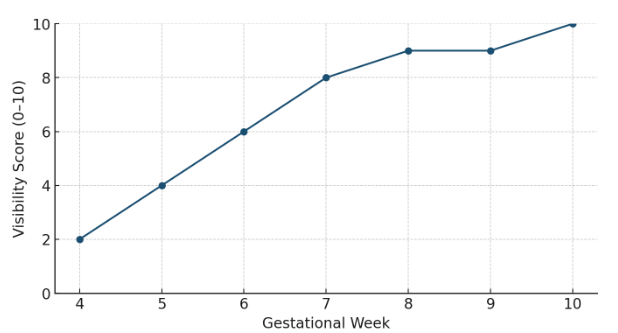
Timeline showing the visibility of early pregnancy structures (gestational sac, yolk sac, fetal pole, heartbeat) via TVS from 4–10 weeks (Rubio et al. 2020).
Abnormal Bleeding or Pelvic Pain
TVS helps identify:
- Endometrial polyps
- Fibroids
- Thickening or irregularities of the uterine lining
Fertility Assessment
TVS allows clinicians to evaluate:
- Follicle development
- Ovaries
- Endometrial readiness for implantation
- Structural issues affecting conception
Ovarian Cysts and Other Pelvic Conditions
TVS can differentiate various types of ovarian cysts and masses, supporting accurate diagnosis and management.
TVS Scan vs. Trans-Abdominal (TAS) Scan
Both scans are valuable, and doctors often use them together for a complete assessment.
Feature | Transvaginal Ultrasound (TVS) | Abdominal Ultrasound (TAS) |
|---|---|---|
Image clarity | Higher resolution | Less detailed |
Best for | Early pregnancy, endometrium, ovaries | Large pelvic masses, overall mapping |
Limitations | Mild discomfort | Requires full bladder |
Body habitus effect | Reliable in obesity or bowel gas | May be limited |
Role in care | Close-up imaging | Complements TVS |
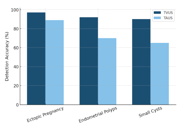
Chart showing diagnostic accuracy of TVS and TAS in detecting ectopic pregnancy, endometrial polyps, and small ovarian cysts (Hu et al. 2023).
Patient Comfort and Consent
Because TVS is an intimate scan, informed consent and patient comfort are essential. Patients receive a clear explanation of the procedure, may ask questions at any time, and can decline or stop the scan if uncomfortable. A trained female chaperone is always present. A protective probe cover is used for hygiene and privacy.
Safety and Infection Control
Every transvaginal probe undergoes strict high-level disinfection after each use. This removes bacteria, viruses, and other pathogens. Single-use probe covers and controlled gel application further enhance safety.
These practices follow international and Malaysian guidelines to ensure safe, reliable care.
What to Expect During a TVS
A TVS typically takes 5–10 minutes.
Steps include:
- Changing into a gown and lying comfortably on the examination bed
- Gentle insertion of a slim, covered probe.
- Real-time imaging and explanation by the clinician
- Removal of the probe and completion of the scan
Most patients experience only mild discomfort, and there is no downtime afterward.
Why Many Women Choose TVS at Metro IVF
- High-resolution imaging for accurate diagnosis
- Early and reliable pregnancy assessment
- Comprehensive fertility evaluation
- A private, respectful, patient-centred environment
TVS helps doctors make confident, timely decisions that support women’s health and fertility outcomes.
